
Application Note
Live cell kinetics assay utilizing the ImageXpress Micro System
- Monitor time-course experiments for hours or days
- Return to the exact same FOV every time
- Create videos with only two clicks
Introduction
The ImageXpress® Micro Confocal/ XLS Widefield High-Content Imaging System from Molecular Devices includes optional Fluidics and Environmental Control modules, enabling single-channel pipetting for compound addition and media exchange during rapid kinetics or extended time-lapse experiments. The Fluidics option is powered by MetaXpress® High Content Image Acquisition and Analysis Software, allowing precise control of each pipetting event. Each system uses 96-well and 384-well format pipette tips for accurate dispensing of volumes between 3 and 200 µL. Two compound plates or media reservoirs can be loaded onto the system with optional heating. The ImageXpress Micro System equipped with Environmental Control and Fluidics modules enables scientists to add compounds and then monitor living cellular responses over a time-span of minutes or even days.
The Transfluor® Cell-Based GPCR Assay Kit is a valuable tool used during high content screening (HCS) to track G-protein coupled receptor (GPCR) activation by quantifying GPCR desensitization and recycling using GFP-labeled ß-arrestin with automated image analysis. The Transfluor Assay is a widely accepted mode of GPCR screening that has been validated on over 90 different GPCRs, and is used as both a primary and secondary screening method in pharmaceutical drug discovery programs. It monitors the “universal” GPCR desensitization and recycling pathway which is independent of downstream G-protein signaling and can thus be used to study nearly all GPCRs. It requires the over-expression of the gene for the GPCR of interest in a cell line expressing the GFPtagged ß-arrestin gene.
Each Transfluor Assay utilizes image analysis to quantify the internalization of GFP-tagged ß-arrestin associated with a GPCR of interest following its activation. Unstimulated cells display diffuse cytoplasmic ß-arrestin-GFP fluorescence. Upon activation of the GPCR, ß-arrestin targets the receptor for internalization, resulting in the appearance of small fluorescent clathrin-coated pits. If the receptor has a high affinity for ß-arrestin, the cells subsequently form fluorescent endocytic vesicles, which are larger and brighter than pits (Figures 1 and 2).
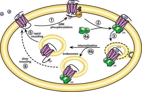
Figure 1. Transfluor Assay technology. Role of arrestins in model of GPCR desensitization and resensitization. (1) Agonist-activated GPCRs are phosphorylated by GRKs (G-protein coupled receptor kinases) on their carboxyl-terminal tails. (2) Arrestins (Arr) translocate to and bind the agonist-occupied, GRK-phosphorylated receptors at the plasma membrane. (3) Arrestins target the desensitized receptors to clathrin-coated pits for endocytosis. (4a) Some receptors bind arrestin with low affinity and dissociate from arrestin at or near the plasma membrane. (5) These receptors internalize without arrestin into endocytic vesicles and recycle rapidly. (4b) Other receptors bind arrestin with high affinity and remain associated with arrestin such that the receptor-arrestin complex internalizes as a unit into endocytic vesicles. (6) These receptors recycle slowly.
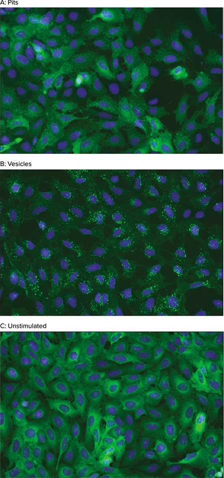
Figure 2. Transfluor Assay cellular phenotypes. Activation of GPCRs in the Transfluor Assay induces the formation of ß-arrestin-GFP-containing pits (A) or vesicles (B). Unstimulated cells show diffuse cytoplasmic distribution of ß-arrestin-GFP (C). Images were acquired at 20X magnification on an ImageXpress Micro System. Nuclei (blue) were stained with Hoechst 33342.
The Transfluor Assay is traditionally an endpoint assay with the agonist mode being quantified after a 60-minute induction with the agonist. In the antagonist mode, measurement is made after a 45-minute antagonist induction and subsequent 60-minute agonist induction. In the presence of compound the induction often occurs over a several minute time scale and eventually formation of pits achieves a steady state. This allows a convenient and robust endpoint “fix and stain” approach.
This experiment was designed to show the kinetics of the Transfluor Assay response to agonist and antagonist and to determine if the 60-minute time point was optimal for acquiring images in an endpoint assay. The experiment was facilitated using the environmental control chamber and scheduled fluidics addition on the ImageXpress Micro System.
With the addition of the Fluidics module to the ImageXpress Micro System, kinetic experiments can be used to study the formation and regression of resulting pits and vesicles in the Transfluor Assay. The MetaXpress software fully automates both the image acquisition and analysis of the resultant image data. The Transfluor Application Module was specifically designed and optimized for analyzing images from Transfluor Assays. Currently custom journals using the Transfluor module are used to analyze kinetic Transfluor Assay data.
Materials
Practice of Transfluor Assays requires a license from Molecular Devices. Transfluor Assay reagents are available to Transfluor Assay licensees from Molecular Devices. Contact Molecular Devices for details.
Note: All solutions should be freshly prepared immediately before each experiment.
- ß-arrestin-GFP and ß-2-adrenergic receptor (ß2AR)-expressing U2OS cells (Molecular Devices)
- Costar 96-well microplates, poly-D-lysine coated, clear bottom, black polystyrene (Corning Cat. #3667). Used for cellular assay plate.
- Costar 96-well clear microplates, round bottom polypropylene, sterile (Corning Cat. #3359). Used for source/compound plate.
- U2OS culture media: MEM (Gibco Cat. #11095) with 10% heat-inactivated FBS (Gibco Cat. #10082), 10 µg/mL gentamicin (Gibco Cat. #15710), 10 mM HEPES (Gibco Cat. #11344), 0.4 mg/mL Zeocin (Gibco Cat. #R250), and 0.4 mg/mL G418 (Gibco Cat. #10131)
- Starvation media: Serum-, phenol red-, and antibiotic-free MEM (Gibco Cat. #51200) with 10 mM HEPES
- Ascorbic acid media: 1 mg/mL solution of ascorbic acid (sodium L-ascorbate, Sigma Cat. #A7631) in Starvation media
- Isoproterenol stock solution: 5 mM stock solution of (-)-isoproterenol (+)-bitartrate salt (Sigma Cat. #I2760) in cell-culture grade DMSO (Sigma Cat. #D2650)
- Propanolol stock solution: 5 mM stock solution of (s)-(-)-propanolol hydrochloride (Sigma Cat. P8688) in cell-culture grade DMSO (Sigma Cat. #D2650)
- DPBS, calcium chloride- and magnesium chloride-free, (Sigma Cat. #D8537)
- 4% methanol-free formaldehyde: 16% methanol-free formaldehyde (Polysciences Cat. #18814-20) diluted in DPBS with calcium chloride and magnesium chloride (Gibco Cat. #14040)
- Hoechst 33342, 10 mg/mL (Invitrogen Cat. #H3570)
Methods
Assay protocol
Step 1. Plate cells at 35,000 cells/well in 100 µL of U2OS media in a poly-D-lysine coated 96-well microplate. Incubate overnight at 37°C, 5% CO2.
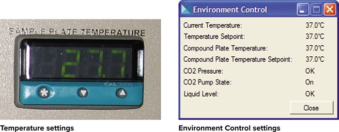
Step 2. Turn on ImageXpress Micro System and use Options Control box to set the temperature on the compound and sample plate positions to 37°C. Check CO2 supply from 5% CO2/95% air tank. Allow system to equilibrate for at least 1 hour.
Step 3. Aspirate media and replace with 75 µL of Starvation media containing a 1:300,000 dilution of Hoechst 33342. Protect from light and incubate at 37ºC, 5% CO2 for 20 minutes. Set up the instrument parameters during the incubation (see the section “ImageXpress Micro Assay Setup” below).
Step 4. Aspirate Hoechst stain and replace with 75 µL of fresh Starvation media.
Step 5. Load plate into ImageXpress Micro System with the Environmental Control (EC) gasket.
Step 6. Image each well at 1-minute intervals for 62 minutes, starting at t=1 minute.
Step 7. Utilizing the ImageXpress Micro System, add a new event to add 75 µL of agonist or control at t= 2 (after imaging) from compound plate 1. Mix 1 time, exchanging the pipette tip after the mix step.
a) Well A01-A03: Control, Starvation media only (no FBS)
b) Well B01-B03: 2X Isoproterenol (final concentration 100 nM) in Ascorbic acid media
c) Well C01-C03: 2X Isoproterenol (final concentration 100 nM) in Ascorbic acid media
Step 8. Add a new event to wash out wells with 75 µL antagonist or Starvation media at t=32 from compound plate 2. (3 exchanges, 50% exchange, new tip each exchange)
a) Well A01-A03: Control, Starvation media supplemented with 10% FBS
b) Well B01-B03: 100 nM Propanolol in Starvation media
c) Well C01-C03: Starvation media supplemented with 10% FBS
ImageXpress Micro System assay setup
During the 20-minute serum starvation and nuclear staining incubation period, set up the instrument parameters. The ImageXpress Micro System must have the Environmental Control and Fluidics option installed.
Step 1. Verify environmental chamber has stabilized by selecting Devices / Environmental Control to view the current status of the environmental control chamber.
- Compound and sample plate temperatures should read 37°C.
- If the CO 2 pressure is at the appropriate level, it will read OK.
- If there is sufficient liquid in the humidity chamber, the Liquid Level will read OK.
Step 2. Under Plate Acquisition Setup/ Fluidics tab, select Configure Stations and then System Properties to define the following parameters.
- Draw and Dispense speed: 5 µL/sec
- Post draw air gap: 2 µL
- Pre draw air gap: 10 µL
- Wet dispense: ON
- Smart dispense: OFF
- Draw overfill: 0 µL
- Pump settle time: 100 ms
Within the Configure Stations dialog select:
- Track volume: ON
- Track liquid surface: ON
- Initial volume for tracking for compound plates: 150 µL
- Under Plate Acquisition Setup/ Fluidics tab, in Scheduled Events:
- After Image 2, Add compound, 75 µL, Compound Plate 1, all selected wells, mix 1X, new tip each exchange
- After Image 32, Washout, 75 µL, Compound Plate 2, all selected wells, with tip exchange, 3 exchanges
Step 3. Prepare compound plates 1 and 2: (MetaXpress software performs 1-to-1 mapping of the microplate wells)
- Compound plate 1: Dispense 150 µL of 2X 200 nM Isoproterenol in Ascorbic acid media (Final concentration 100 nM) into wells B01-C03 and 150uL Starvation media in a Costar 3904 microplate into wells A01-A03.
- Compound plate 2: Dispense 275 µL of 1X Propanolol in Starvation media in Co-star 3904 microplate into wells B01-B03 and Starvation media supplemented with 10% FBS into wells A01-A03 and C01-C03.
Image acquisition
Using MetaXpress software, acquire 2x2 binned images with camera gain set to 1 using DAPI and FITC filter sets and a 20X Plan Fluor ELWD (NA = 0.45) objective. The results described here were obtained from single site per well acquisitions.
Reviewing images
In MetaXpress software, load your experiment through the Review Plate Data dialog box under the Screening menu and choose Select Plate to navigate to your specific plate. Once the experiment is loaded, the selected wells will be displayed on the montage (Figure 3A). Click on one of the wells to display the full-resolution image(s) (Figure 3B). Images may be viewed as single wavelengths or as a color composite (as shown in the three following figures).
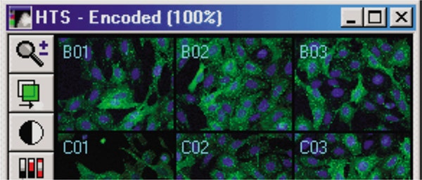
Figure 3A. Transfluor Assay experimental image montage.
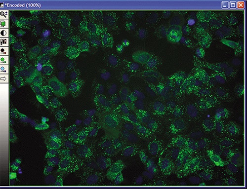
Figure 3B. Selected well data.
To view individual images, either change the Timepoint on the Review Plate Data dialog box, or change the view to Time vs. Well (recommended for kinetic experiments). Right-click on an individual well to select it, then click on Load Images to load all timepoints into a stack, which may be viewed and exported as a movie (Figure 3C).
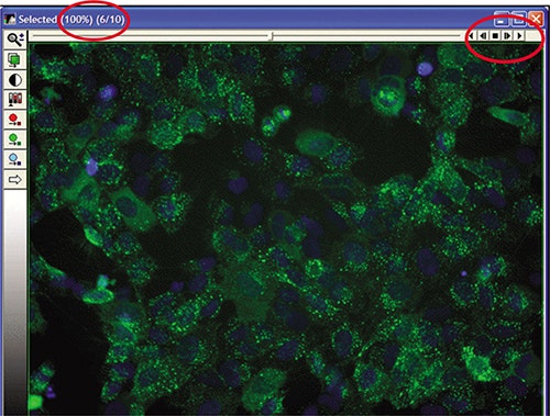
Figure 3C. Stack of timepoint data.
Image analysis
Kinetic data can be analyzed with the Kinetic Transfluor Analysis journals. Visit the Knowledge Base section of the Molecular Devices website and follow the detailed instructions to download, install and run the kinetic analysis journal.
If assay conditions change, the analysis parameters in the journal may need to be re-optimized. Use the instructions from Article 19736 on the Knowledge Base site to determine the correct Transfluor Assay settings.
Results
U2OS cells expressing ß-arrestin-GFP and either wild-type ß2AR (pit-formers) or enhanced ß2AR (vesicle formers) were treated with assay media only, or Isoproterenol (agonist) plus or minus propranolol (antagonist). The enhanced ß2AR has a modified C-terminal tail which enables a greater affinity for ß-arrestin and allows fluorescent vesicle formation.
Images were acquired on an ImageXpress Micro System equipped with the Environmental Control and Fluidics options as described in the Materials and Methods, and analyzed with custom Kinetic Transfluor Analysis journals.
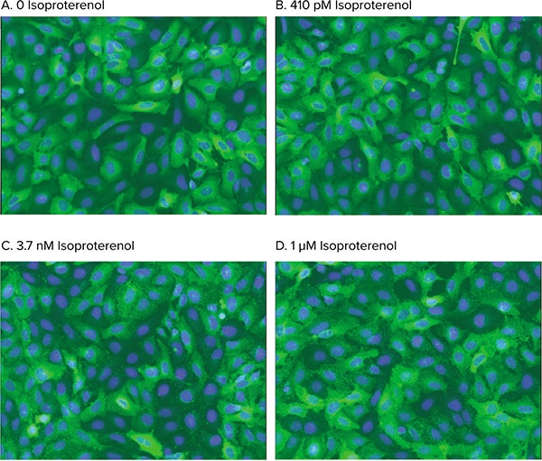
Figure 4. Agonist stimulation of pit formation. Analyzed images of dose-dependent ß-arrestin-GFP translocation upon activation of WTß2AR. Cells were treated with 0 (A), 410 pM (B), 3.7 nM (C), or 1 µM (D) Isoproterenol. The Transfluor Assay Application Module accurately identified pit formation induced by agonist stimulation.
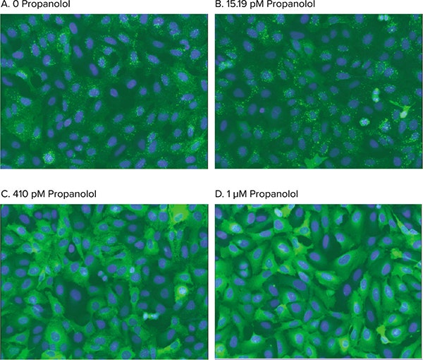
Figure 5. Agonist stimulation of vesicle formation. Analyzed images of dose-dependent inhibition of arrestin-GFP translocation caused by stimulation of a tail-modified ß2AR. Cells were treated with 0 (A), 15.19 pM (B), 410 pM (C), or 1 µM (D) propanolol and stimulated with 50 nM Isoproterenol. The Transfluor Assay Application Module accurately identified vesicle formation blocked by antagonist inhibition.
Dose-response and dose-inhibition analysis
In both pit- and vesicle-forming cell lines, the time-dependent responses of the agonist Isoproterenol and the antagonist propranolol were measured with the custom journals based on the Transfluor Assay Application Module within the MetaXpress software. Each dose was run on a single well across 4 replicate experiments.
Using AcuityXpress™ Software, Hill curve fitting was used to determine EC50 and IC50 values based on pit or vesicle area per cell, plotted for each timepoint in the time course.
The data displayed is for one single time course experiment. The ß2AR-enhanced cells were treated with three conditions: basal, agonist followed by antagonist, and agonist only. The ß2AR-wild type cells were treated with two conditions: basal and agonist only.
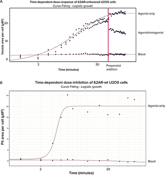
Figure 6. Dose-response and dose-inhibition curves. A. ß2AR-enhanced cells with basal, agonist/ antagonist treatment, and agonist-only). B. ß2AR-wild type cells with basal and agonist-only treatment.
Conclusion
The ImageXpress Micro System equipped with the Environmental Control and Fluidics option module successfully executed a kinetic Transfluor Assay. The MetaXpress software kinetic analysis journal also clearly identified cells and measured pits and vesicles formed by the ß-arrestin-GFP translocation to activated GPCRs in the Transfluor Assay. Graphs displaying data analyzed with AcuityXpress Software show that the responses are stable and wellseparated if a 60-minute endpoint read is acquired. These results demonstrate that the ImageXpress Micro System equipped with the Environmental Control and Fluidics Module in conjunction with the Transfluor Assay is a robust tool in conducting kinetics experiments in screening GPCR modulators. To view the time-lapse movies of the vesicle-former responses, please visit our web site
References
- Oakley, R.H., Hudson, C.C., Cruickshank, R.D., Meyers, D.M., Payne, R.E. Jr., Rhem, S.M., Loomis, C.R. The cellular distribution of fluorescently labeled arrestins provides a robust, sensitive, and universal assay for screening G-protein-coupled receptors. Assay Drug Devel Tech 2003; 1:21-30.
- Oakley, R.H., Laporte, S.A., Holt, J.A., Barak, L.S., Caron, M.G. Association of beta-arrestin with G-protein-coupled receptors during clathrin-mediated endocytosis dictates the profile of receptor resensitization. J Biol Chem 1999; 274:32248-32257.
- Catalano, S.; de Bruin, S.; Bosworth, J.; Gliksman, N.; Best, K.; Rickert, P. HighContent screening of GPCR activation with MetaXpress, AcuityXpress and the Transfluor assay system. Molecular Devices Application Note.
Learn more about ImageXpress Micro Confocal High-Content Imaging System >>