
Videos & Webinars
Unleash the power of Cell Painting
The Science Explorer interviews Angeline Lim, an Applications Scientist at Molecular Devices, to learn more about Cell Painting and the impact it is having on cell biology research and drug discovery.
Watch the video to view the full interview. See a snippet of the interview below.
Tiffany:
Can you tell us what Cell Painting is?
Angeline:
Sure, so Cell Painting essentially is a high-content, image-based assay that you would use for morphological profiling. In a Cell Painting assay you would use up to six fluorescent dyes to label different components of the cell. That would include the nucleus, the endoplasmic reticulum, mitochondria, cytoskeleton, and more. The idea is to paint as much of the cell as possible so you can get a representative image of the cell. And then from these images, you would use automated image analysis to extract over 100-1000 parameters of the cell. With these measurements, you can get intensities, texture, shape, and even the proximity to neighboring structures. So in this way, you can get an idea of the spatial relationship between organelles or even within the same group of organelles that you're studying. All of these measurements form what we refer to as the phenotypic profile.
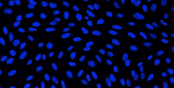
Nucleus
Dye: Hoechst 33342
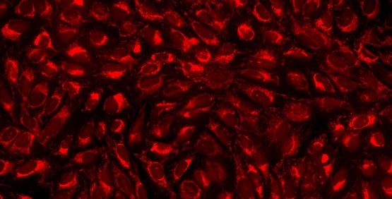
Mitochondria
Dye: MitoTracker Deep Red
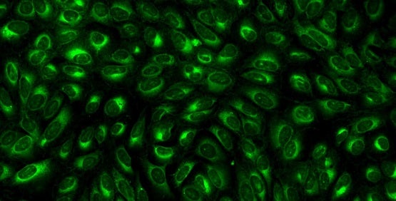
Endoplasmic reticulum
Dye: Concanavalin A/Alexa Fluor 488 conjugate
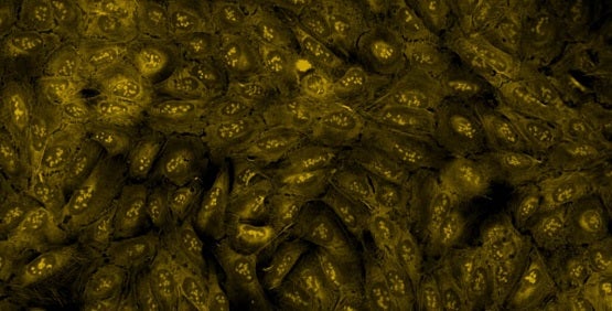
Nucleoli, cytoplasmic RNA
Dye: SYT0 14 green fluorescent nucleic acid stain
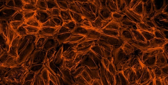
F-actin cytoskeleton, Golgi, plasma membrane
Dye: Phalloidin/Alexa Fluor 568 conjugate, wheat- germ agglutinin/Alexa Fluor 555 conjugate
Tiffany:
So, can you walk us through the technique in a bit more detail?
Angeline:
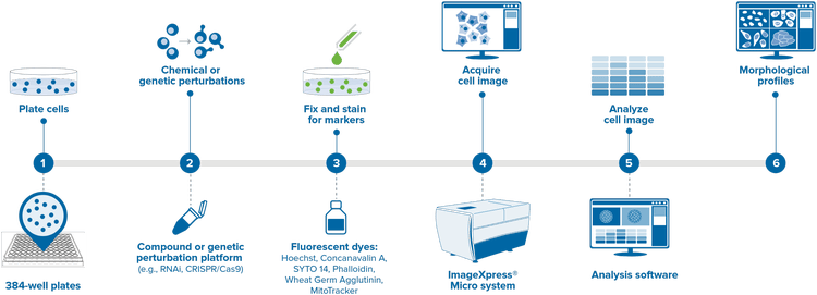
Well, what you start with is of course a multi-well plate. You put your cells in these plates. Next, you introduce some kind of perturbation. It can be chemical or genetic. With chemical perturbations, you're doing, for example, a drug screen. With genetics you could, for example, use RNAi to help understand the function of specific genes. Following that, you would apply the set of Cell Painting dyes so that you stain and label all these different parts of the cell, and then you take your plate, and put it in a high-content imager, such as our ImageXpress® Micro High-Content Imaging System. After you have these images, you would run automated image analysis, which identifies individual cells and the sub-components within these cells. You would measure and extract features to derive the rich morphological profile, which will be suitable for detecting subtle changes in phenotypes.
Tiffany:
What would you say is the advantage of using Molecular Devices high-content imaging systems in particular for Cell Painting?
Angeline:
I would say there are many, but some things to highlight would be that you could use any of our ImageXpress Micro systems in the imaging step for your Cell Painting assay. Actually, the pioneering work on Cell Painting came from the Broad Institute. They used our ImageXpress system to develop a Cell Painting assay. The dyes were optimized for the filter sets we have in our system. And so at this point, I would say that having one of our ImageXpress systems is almost a plug and play. You just have to prep the plate, put it on the instrument and read it.
We are also constantly working to improve our products. We add new features or have new modules that will be beneficial for our customers. Right now for Cell Painting, one of the bottlenecks is in imaging speed, because you are getting images from a 384-well plate. Within each of these wells, most people would image nine sites in each well. Multiply this by five channels and you get a huge amount of data. And so one way to improve the imaging speed would be to use a "larger" (high-resolution) camera that would give you the same amount of data when you image fewer sites. You will get a larger field of view with a lower magnification objective and analyze the images. In this way, you can read the entire plate faster, without sacrificing image quality.