Application Note
Using the NanoOrange Protein Kit with SpectraMax Microplate Readers
- Greatly improved sensitivity over absorbance methods
- Wide dynamic range from 10 ng/mL to 10 μg/mL
- Easy data acquisition and analysis with pre-configured SoftMax Pro Software protocol
Introduction
This application note describes how to use the NanoOrange® Protein Quantitation Kit from Life Technologies in SpectraMax ® microplate readers with the fluorescence detection mode and SoftMax ® Pro Software from Molecular Devices. This assay is much more sensitive than traditional photometer methods, such as absorbance 280, BCA, Bradford or Lowry assays. The dynamic range of this assay in microplate format is 10 ng/mL to 10 μg/mL. The data presented in this application note confirm the dynamic range and lower detection limit described in the manual for the NanoOrange Kit (MP 06666).
Materials
- NanoOrange Protein Quantitation Kit from Life Technologies (catalog number N-6666)
- Protein for standard curve (BSA is supplied with the kit)
- Distilled water
- Heating block at 90-96°C
- Plastic tubes (1 mL microfuge tubes)
- Solid black microplates
- Gemini XPS Microplate Reader (or other SpectraMax reader with fluorescence detection mode)
- SoftMax Pro Software
Methods
Set up the instrument and software
Step 1: Turn on your microplate reader.
Step 2: Launch SoftMax Pro Software. Open the Protocol Library, locate the ‘Protein Quant’ folder, and open the pre-configured protocol ‘NanoOrange’.
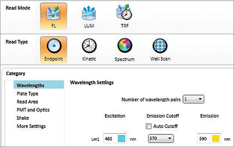
Step 3: Click “Settings” and a screen will appear (Figure 1). First choose Read Mode ‘FL’ and Read Type ‘Endpoint’, and then select wavelengths: Excitation 485 nm, Emission Cutoff 570 nm, and Emission 590 nm. Continue down the Category panel at the left side of the Settings window to select parameters appropriate to the assay plate type, wells to read, etc. For PMT and Optics, Automatic PMT Gain is recommended. The number of flashes per read should be set to at least 20 (more flashes may offer greater precision but will lengthen the read time).

Figure 1. Plate reader settings for NanoOrange assay. Typical settings for SpectraMax readers with fluorescence detection mode are shown.
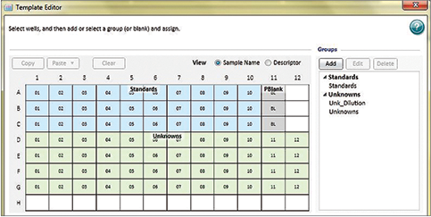
Step 4: Use the Template Editor to assign wells to ‘Standards’ and ‘Unknowns’ groups (Figure 2). This enables the standard curve to be plotted and sample concentrations to be calculated automatically by SoftMax Pro Software. Be sure to include a plate blank.

Figure 2. Template setup. Assigning wells to ‘Standards’ and ‘Unknowns’ groups in the Template Editor enables automatic plotting of the standard curve as well as data analysis.
Prepare the assay
The method for this assay follows the instructions in the product information sheet for NanoOrange, except the assay volume is proportionately reduced from 2.0 mL to 180 μL to fit the 96-well microplate format.
Step 1: Dilute the concentrated Component B (NanoOrange protein quantitation diluent) 1:10 in distilled water (one part Component B to nine parts distilled water).
Step 2: Dilute the concentrated Component A (NanoOrange protein quantitation reagent) 1:500 with the working concentration (1x) NanoOrange protein quantitation diluent to make the NanoOrange working solution.
Note: This should be done just prior to the experiment (the working solution should be used within a few hours of preparation). Only prepare enough of the NanoOrange working solution for the number of samples (including standards, unknowns, blanks and any other controls) in the assay.
Protect the NanoOrange working solution from light by placing it in an opaque bottle, by wrapping a non-opaque container in aluminum foil, or by placing the solution in a dark cupboard or drawer. It is important to protect the working solution from light to prevent photodegradation of the reagent.
Step 3: Dilute 2 mg/mL BSA in NanoOrange working solution to make 10 μg/mL and 1 μg/mL stock solutions.
Step 4: The reference curve and unknowns are prepared using the NanoOrange working solution. The standard curve shown in this application note was prepared using a 10 μg/mL and 1 μg/mL stock of BSA. The volumes given in this table are enough to prepare 5 replicates of each concentration. You may want to adjust the volumes, depending on the number of replicates you want to run. Mix the appropriate BSA with the NanoOrange working solution into 11 brown (for protection from light) microfuge tubes as described in Table 1.
Final BSA Concentration
(μg/μL)in assay
NanoOrange Working
Solution (μL)
Table 1. Preparation of BSA standards.
Step 5: Incubate samples at 95°C for 10 minutes (protected from light), then cool to room temperature (approximately 20 minutes), again keeping samples protected from light.
Step 6: Transfer 180 μL of each sample to the appropriate well of the microplate.
Triplicates of each BSA concentration were made for this assay plate.
Read the microplate
Step 1: Place the microplate in the reader.
Step 2: Click the Read button in SoftMax Pro Software. The microplate reader will read the plate and the relative fluorescence units will be displayed in the Plate section.
Analyze the data
Step 1: After the microplate has been read, the RFUs will be displayed in the Plate section. The data will be analyzed in the Group tables that you created while setting up the template (see the section “Setting up the instrument and software” earlier in this document).
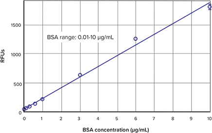
Step 2: A standard curve will be plotted automatically, as long as the pre-configured protocol is used and the appropriate wells are assigned to the Standards group in the Template Editor. Choose the appropriate curve fit from the drop-down Curve Fit menu in the graph section’s tool bar. When plotting the standard curve shown in Figure 3, a linear curve fit was used.
Results
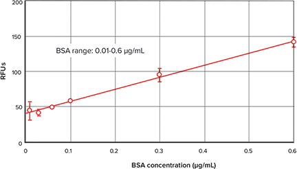
Figure 3 shows the standard curve obtained using the NanoOrange Protein Quantitation Kit with the BSA standard provided in the kit. The low end of the standard curve is shown in Figure 4. Similar data and curves are produced using other SpectraMax readers with fluorescence detection mode (data not shown).

Figure 3. NanoOrange standard curve. Standard curve obtained using BSA standards and a linear curve fit.

Figure 4. Standard curve low end detail. Detail of the lower end of the standard curve shown in Figure 3.
Conclusion
The NanoOrange assay, when run on a Gemini XPS Microplate Reader or other SpectraMax fluorescence microplate reader with SoftMax Pro Software, is a quick, sensitive detection method for proteins. The analysis capabilities of SoftMax Pro Software provide quantitation in an easy-to-read, user-customizable report format.