SCIENTIFIC POSTER
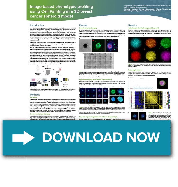
Image-based phenotypic profiling using Cell Painting in a 3D breast cancer spheroid model

Most potential oncology drugs fail the drug development pipeline, despite having promising data for their efficacy in vitro.
Here we demonstrate a screen using patient-derived 3D spheroids (tumoroids) where—in addition to readouts on cell viability—we adapted the Cell Painting assay for the 3D tumor model. Tumoroids were formed from primary cells isolated from a patient-derived tumor explant that represents metaplastic breast cancer with a triple-negative breast cancer. Results demonstrate the feasibility of using the Cell Painting assay on 3D cell models.
Heading
Register to download your scientific poster today
Product family
Imaging
Product primary application
ImageXpress Confocal HT.ai; MetaXpress High-Content Image Acquisition and Analysis Software; IN Carta Image Analysis Software; StratoMineR
CMP
7014u000001AQ9vAAG
Title
Image-based phenotypic profiling using Cell Painting in a 3D breast cancer spheroid model
Resource URL
Resource Image URL
