
Application Note
Improved productivity of cell line development using fully integrated automated solutions: a series of case studies with the CloneSelect Imager
- Ability to scale up on throughput capabilities as needs change and grow
- Significantly reduce redundant manual operation and increase throughput of your research by providing walk-away time, overnight operation and full workflow automation
- More consistent processing of plates provides more reliable data
- Better environmental control for cell maintenance during assays
Introduction
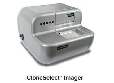
Cell line development requires screening tens of thousands of clones in order to find the few clones that are stable, grow well, and produce high yields of bioproduct. In addition to finding these rare clones, researchers must show that their bioproduct of interest originated from a single clone (i.e. monoclonal) in order to pass FDA regulations. Thus, scientists are often tasked with the time-consuming and tedious process of screening numerous clones on a cell-by-cell basis. In order to significantly reduce this burden, Molecular Devices has developed the CloneSelect™ Imager, an automated imaging system that is able to capture images of single cells and monitor their growth over time. Here we present three case studies in the order of increasing throughput, where we have integrated the CloneSelect Imager with an automated workflow in order to significantly improve workload capacity.
Case study #1 highlights a simple automated solution designed to alleviate the burden of manually screening thousands of clones over the course of one day. Case study #2 illustrates our ability to extend the time frame for automating the monitoring of clones for as long as desired (typically 2-3 weeks). Finally, case study #3 provides an overview of how to combine the monitoring of clones with solutions for assessing the quality of bioproduct produced.
Case Study #1 – Automated delivery of 96-well microplates to cell colony imager
Instrument Setup: CloneSelect Imager + robotic arm + plate hotel
While there are many strategies available to help show proof of monoclonality, providing images of single cells following limiting dilution has played a critical role in this process because it is relatively easy to do and provides fairly definitive evidence. However, capturing an image of a single cell for the thousands of clones that must be screened can be an extremely tiresome process, especially if done with a manual microscope. Thus, the CloneSelect Imager was developed to provide rapid imaging of entire multi-well plates in order to assess whether each well contains a single cell and to further monitor growth over time. (Please see “Rapid monoclonality verification methods to boost cell line development”). As a stand-alone instrument, the CloneSelect Imager can only image one plate at a time though, requiring users to manually load plates into the system.
However, by fully integrating the CloneSelect Imager with a third-party robot that can handle 48 plates at room temperature during a single run (Fig. 1), the walk-away time for of our customer has increased significantly by over 2 hours.
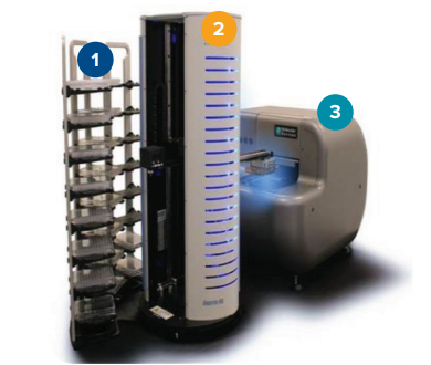
Figure 1. A typical instrument setup to achieve automated plate delivery to the cell colony imager. 1. Plate hotel. 2. Robotic arm. 3. CloneSelect Imager.
Case Study #2 – Automated delivery of 96-well microplates from an incubator to cell imager and back
Instrument Setup: CloneSelect Imager + robotic arm + incubator
Although case study #1 certainly increased the walk-away time for our customer on a day-to-day basis, setup still requires loading and unloading of plates from the plate hotel everyday. Furthermore, plates remain at ambient temperature over the course of the run, which may not be ideal for long-term viability. In this case, we mitigated the issue of long-term viability by integrating the CloneSelect Imager with an incubator using a robotic arm (Fig. 2). Not only did this reduce temperature variability, our customer was also able to walk away from the experiment for 2-3 weeks without having to touch the plates, thus increasing their productivity while also reducing contamination and other variability from plate handling. In this example, we highlight integration with a 42-plate incubator, but we have also provided a solution for a large capacity incubator housing approximately 200 plates.
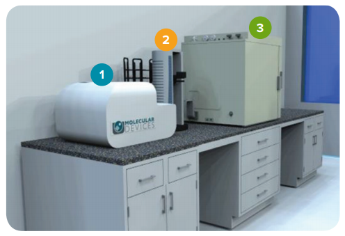
Figure 2. A typical instrument setup to achieve automated plate delivery between the cell colony imager and incubator. 1. CloneSelect Imager. 2. Robotic arm. 3. Incubator.
Case Study #3 – Automated delivery of 96-well microplates from an incubator to the CloneSelect Imager, followed by automated transfer of supernatant to plates for downstream analysis
Instrument Setup: CloneSelect Imager + robotic arm + incubator + liquid handler
The next step after monitoring the growth of single cells into colonies is to assess their productivity. This can be accomplished in a number of ways, including detection of antibodies by ELISA using a plate reader, such as the SpectraMax® i3x reader from Molecular Devices, or by using label-free methods, such as the Octet® System from ForteBio.
Often, this requires that supernatant be pulled from those clones that grew into well-established colonies, but the process can be challenging to perform manually since clones grow at different rates. Here we highlight an automated solution whereby single cells deposited into multi-well plates were loaded into an incubator and monitored for growth on the CloneSelect Imager over the course of 2 weeks (Fig. 3). During this 2-week period, wells with a confluency of >80% were automatically selected for downstream analysis, which allowed for the different growth rates of clones. Supernatant from flagged wells was aspirated through the use of an automated liquid handler provided by Beckman Coulter and deposited into multi-well plates ready for assessment by ELISA.
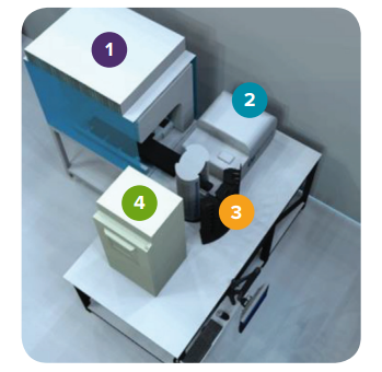
Figure 3. A typical instrument setup to achieve automated plate delivery between the cell colony imager, the incubator, and liquid handler. 1. Liquid handler. 2. CloneSelect Imager. 3. Robotic arm. 4. Incubator.
Summary
With the ever-increasing demand to shorten timelines for cell line development, automated solutions such as those shown in these three case studies provide the necessary walk-away time, increased throughput, and reliability required for moving antibody candidates through the cell line development pipeline (Table 1). In addition to the higher throughput provided, it is imperative that customers walk away with the assurance that reliably high-quality data is captured and documented.
• Free up manual labor
• Customer can enjoy a walk-away time of 2 hours
• Can automatically process 48 plates during a single run
• Free up manual labor
• Customer can enjoy a walk-away time of 2-3 weeks
• Reduce contamination and variation from plate handling
• Unlimited potential for processing of plates (only depends on the capacity size of the incubator)
• Free up manual labor
• Customer can enjoy a walk-away time of 2 weeks
• Reduce contamination and variation from plate handling
• Automated collection of supernatant from colonies on different days (in order to compensate for variable growth rates) for further downstream analysis
Table 1. A summary illustrating the benefits of each customized automated workflow.